The title should read “Externally rotate the femurs (if your femurs are internally rotated) and align your pelvis to support your uterus,” But that would be too long a title.
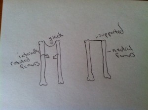
An oversimplified explanation of how the femur position affects the pelvic floor and uterus. Even the feet collapse in (pronate) when the femurs are internally rotated. I can actually feel my organs being dragged downward!
Slack muscles will respond by shortening, but short doesn’t equate to strong. In fact, muscles that have shortened are weaker. This can be demonstrated on an EMG.
Ligaments of the uterus and ovaries aren’t just for support, but they also are pathways for blood, lymph, and nerve flow to and from the reproductive organs. It’s important to remember, in order for the femur, sacrum, and pelvis to be neutral, the connecting muscles need to be at their correct length. If the muscles aren’t at the correct length, blood, lymph, and nerve flow are negatively affected.
Here is a brief overview of the uterine ligaments and their attachment points:
Round ligaments are thin fibro-muscular cords that run from the lateral aspect of the fundus of the uterus and travel anterolaterally through the inguinal ring and canal and connect to the superficial perineal fascia at the labia majora. The round ligaments pass through the two layers of the broad ligament. The round ligaments lengthen from 4-5 inches to up to 18 inches or so during pregnancy! The round ligaments help maintain the anteverted position of the uterus.
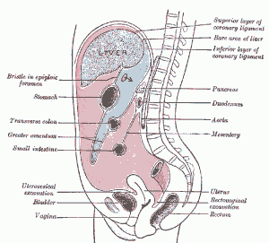
Broad ligament: connects the uterus to the lateral walls of the pelvis. The broad ligament is an extension of the peritoneum and envelops the uterus in its folds, so it appears as two flat wide ligaments extending outward laterally. From the picture below you can see how the position of the rib cage and motility /mobility of the digestive organs can affect the ligaments of the organs below via the peritoneum. Everything is connected!
Utero-sacral ligaments: The name implies that the uterosacral ligaments attach the uterus directly to the sacrum, but that is the case only 7% of the time. This study shows, “the origin of the uterosacral ligament from the genital tract extends from the cervix to the upper vagina. The insertion on the pelvic sidewall occurs to the sacrospinous ligament (see image below) and the coccygeus muscle in 82% of all cases, but in only 7% do the uterosacral ligaments insert on the sacrum and 11% the piriformis muscle, the sciatic foramen, or the ischial spine. This suggests that the uterosacral ligaments exhibit greater anatomic variability than their name implies, and this might be an important insight for the understanding of the pelvic organ support mechanism.” This ligament prevents the cervix from moving forward toward the bladder and from prolapsing. Given the attachment points, it makes sense that the position of the femur, sacrum, and pelvis play a supportive role. Can you see the importance of the squat and posterior push-off (using the gluteus while walking) for optimal sacral positioning? The gluteal muscles are the main force keeping the sacrum from moving anteriorly. Remember the puppeteer image… sacrum=puppeteer handle thingy, uterosacral ligaments=strings, and the uterus=puppet
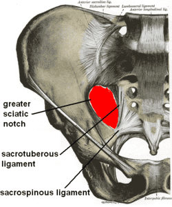
It’s obvious to see how the position of the sacrum and coccyx can affect the uterosacral ligament, and therefore the uterine position
The obturator internus muscle can be accessed through the vaginal wall so pelvic floor PTs who do intravaginal manipulation can be very helpful to women with obturator internus problems. I learned a couple of ways it can be accessed externally in my training in Visceral Manipulation™ and in Maya abdominal therapy. And pelvic/femur/thoracic alignment is very important to the health of these muscles. Issues with the obturator internus can cause vaginal, hip, and back pain, it can also mimic bladder pain, cause painful intercourse upon penetration, and refer pain to the abdomen.
Side note: A very important pelvic nerve called the Pudendal nerve travels through the obturator internus fascia. The pudendal nerve branches are unique to every woman, some have more branches to the mouth of the cervix or to the vagina. Some women have more nerve branches to the clitoris or the perineum. This accounts for different sexual responses in women! No, nothing is wrong with you if you don’t have vaginal orgasms! Something all women and men should know, don’t you think?
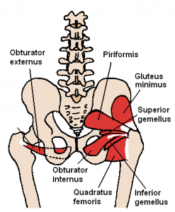
Cardinal ligaments: Are known as the main supporting ligaments of the uterus, upper vagina, and cervix. It attaches in a circular pattern around the cervix and moves laterally to the obturator fascia along the pelvic sidewalls. Inferiorly it is continuous with the fascia on the upper surface of the levator muscles! The obturator internis attaches to the medial aspect of the greater trochanter (see image above), so this leads me to believe that a neutral femur would help support the cardinal ligament. It also, makes me wonder if “incompetent cervix” has to do with a disruption of flow through the ligament to the cervix due to poor alignment or tight pelvic floor.
Yeah, alignment matters! Does it seem natural that we have such a high incidence of uterine prolapse in the US? Our bodies are beautifully designed to hold our organs in place without the help of mesh, pessaries, and 1000 Kegels a day. Recent estimates are around 24% of women in the US have pelvic organ prolapse. I’m guessing it’s even higher because many women with stages one and two don’t seek medical attention. We sit in chairs most of the day, we no longer squat, we wear positive heeled shoes and we don’t use the correct muscles for the most natural task….walking.
Getting Help:
I offer an online Womb Care course where you can learn self abdominal massage and other supportive therapies including pelvic alignment for reproductive health.







So if I have endometriosis that really only developed on my uterosacral ligament – right side – what can that tell me about my alignment?
Sometimes a retroverted uterus can cause endometriosis. Do you know the position of your uterus?
Hi there, thanks for a great post, just clarifying what you mean by ‘rotated femurs’ do you mean walking with toes turned inwards??
Hello Barbara, thank you for another good post. You always know how to convey important information in easy to understand language adding both simplified and detailed drawings. Your contributions are a real asset to Katy’s work. Keep it coming!
Hi Jenny, Great question. No, toes turned inward doesn’t always mean the femurs will be rotated inward. Check out this video for an explanation, http://www.alignedandwell.com/katysays/all-about-your-knees/.
Thanks so much Mona! I really appreciate you taking the time to tell me that.
I have *been told* by a MW that I had a “tilted uterus.” Had three babies with horrifyingly bad sacral contractions. But the MW who delivered my babies did not mention a tilt.
Hi Kris,
The uterus may tilt in several directions. If it was retroverted, it may have corrected itself around 5 months pregnant, sometimes MWs and Drs don’t even mention if the uterus is tilted because they don’t think it is a big deal, or don’t know that there is something that can be done about it.
Well, I went and had Arvigo done anyway! 😉
Great! ATMAT is great for anyone, even if they don’t have a tipped uterus. ATMAT is also beneficial for men, and women who have had hysterectomies. How was your experience with ATMAT?
I agree! It’s amazing how hearing similar information from multiple people totally clarifies things.
And thank you for posting the links to other REs. I’m becoming a total RES-fan girl 🙂
I love reading your posts. You’re great at explaining things and as others have said it is great to read in addition to the Katy Says Blog.
I’m wondering if you also recommend the No More Kegels Course from the Restorative Institute in addition to the Down there for Women for someone who wants more?
Hi Leah, thank you! I absolutely recommend the No More Kegels course. Foot alignment for pelvic floor health is also important, start with the bottom and work up. Search my blog and Katy’s for foot alignment.
Hi!
And thanks for all you do!
I really have a hard time understanding how to correctly rotate my femurs. When I try, I end up on the very outsides of my feet (also heels) and then I have no balance. (And I end up with all my weight in front of me, sort of leaning forwards, because I have to keep my balance somehow…)
And how do you walk without rotating your femurs inwards? (When trying to walk with straight feet)
Hi Hanna,
You can practice getting your femurs out of internal rotation while standing, but I wouldn’t try it while walking until you are able to build your lateral hip strength and other alignment markers (backing the hips up, neutral pelvis, posterior push off, feet ASIS width apart). You need to stand in alignment before you are able to walk in alignment. While practicing getting out of internal rotation during stance, you may start by coming up on the sides of your feet and then practice lowering your toes and ball of foot down. Practice pelvic list and monster walk as well. Once you accomplish neutral femurs in stance without thinking about it, you will start walking in neutral without thinking about it.
Thank you! I will start with the standing then. I tried putting the balls of my feet down, but it’s almost impossible! And it hurts like crazy!
Hello,I have a retroflexed uterus and pelvic organ prolapse. I was given internal vaginal trigger point massages several times, which are excruciating on the R side only. Then I was sent to physical therapy for kegels and hip rotaions…. Next thing I know, I cant walk, cant take care of my family, and have to quit my job. This has been going on for 5-6 months now. I have no fibroids, Im not over weight, before this I could walk all day, and swim and even do cartwheels. The pain is not as bad, but it flares up for days or weeks if I have sex, get my period, or do piriformis exercises. My previous history is strenous jobs, strenous exercising, a R oophorectomy for dermoid and an appendectomy in 2008. I am now considering a hysterectomy, but I dont know for sure. I just want my life back, and Im afraid its not going to happen. I am 45 and in pre menopause. any ideas?? I have no idea how my femurs affect my pelivs, but my R hip is killin me. I even get sciatica now and I never got it before. Thanks!
My cervix tilted to my left about six months ago out of the blue. I’m 38 and have no children. What could have caused this and what should I do about it? I work with weights (squats too!)twice a week (1 hour class at the gym to music) and do pilates and yoga once a week each. I’m very active. My hips tilt forward so I have a bit of a sway back. I recently found out my head is forward a little too.
This answers my question perfectly! I am able to externally rotate my knees while standing, and I am able to do small squats and keep them externally rotated. But as soon as I try to pelvic list or walk, I can’t hold it. I will keep practicing while standing. Thank you!
How would you know just by appearance alone if your femurs are rotated internally or outwardly? Or if they’re really not but only look like they are?
Thanks
by looking at the hamstring tendons as I mentioned in the post. If they had a resting position in external rotation the hamstring tendons would present the opposite way. The hamstring tendons attach to objective boney markers, so that is an objective way to evaluate.
Hello – Do you have suggestions for how to align an anteverted uterus?
Yes! I wrote a post on anteverted uterus.here: https://alignmentmonkey.nurturance.net/2014/bladder-buddies/
and here:https://alignmentmonkey.nurturance.net/2015/anteflexed-uterus/